


AD Biomarkers: Type, Function, Research Applications
Key points
●Alzheimer's disease (AD) biomarkers fall into key biological groups: brain-derived tau (BD-tau), phosphorylated tau (p-Tau181, p-Tau217), amyloid (Aβ40, Aβ42), neuroinflammation (GFAP), and neuroaxonal injury (NfL).
●The AT(N) framework groups biomarkers by the biology they measure, amyloid (A), tau (T), and neurodegeneration (N). The 2024 Alzheimer's Association revised criteria define AD biologically and give blood-based biomarkers a recognized role in research frameworks, but interpretation still depends on the specific biomarker, assay performance, intended use, and validation evidence.
●Vazyme's AD portfolio extends beyond the core markers, including total tau, BD-tau and related tau targets, and additional research biomarkers, with sample-type and species support that varies by assay.
●Blood, CSF, tissue, and organoid samples each carry trade-offs in invasiveness, required sensitivity, and pre-analytical control.
●Assay performance, limit of detection, precision, cross-reactivity, and validated sample compatibility, determines whether your results are comparable with published data.
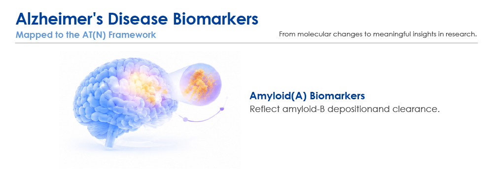
Alzheimer's disease biomarkers are measurable indicators of the biological processes that drive AD: amyloid-β plaque formation, hyperphosphorylated tau aggregation, glial activation, and neuroaxonal injury. For researchers, they answer three practical questions, is the pathology present, is it progressing, and is an intervention changing it?
This guide covers the main AD biomarker types, what each one reflects, which sample types are used in research, and how to build a biomarker panel for your study. It is a research-focused guide and does not cover clinical diagnostic testing.
What Are Alzheimer's Disease Biomarkers?
A biomarker is any measurable indicator of a biological state or process. In AD research, biomarkers stand in for the neuropathological hallmarks of the disease, extracellular amyloid-β plaques and intracellular neurofibrillary tangles of hyperphosphorylated tau, as well as their downstream consequences: neuroinflammation and neurodegeneration.
The field organizes these markers using the AT(N) research framework[1]:
A: Amyloid
Biomarkers of aggregated amyloid-β (e.g., CSF or plasma Aβ42/40 ratio; amyloid PET)
T: Tau
Biomarkers of AD-related tau pathology (e.g., CSF or plasma p-tau; tau PET)
(N): Neurodegeneration
Not specific to AD (e.g., MRI atrophy, FDG-PET, CSF total tau)
The "(N)" is parenthesized deliberately: neurodegeneration also occurs in many non-AD conditions, so it tells you that neural injury is happening, not that AD is the cause.
In 2024, the Alzheimer's Association published revised criteria that define Alzheimer's disease biologically and provide a framework for evaluating biomarkers across the disease continuum[2]. Blood-based biomarkers are an important and evolving part of this field, but their interpretation depends on the specific biomarker, assay performance, intended use, and validation evidence, inclusion in a research framework does not mean that every blood-based assay is suitable for clinical diagnosis. For researchers, the practical takeaway is that plasma biomarkers now sit inside a recognized biological framework, while assay-level validation in your own matrix and species remains a separate, non-negotiable step.
One scope note: clinical diagnosis requires validated, regulated assays. Research-use-only (RUO) assays, including the AD biomarker kits described in this guide, are intended for research applications and are not for use in diagnostic procedures[3].
What Are the Main Types of Alzheimer's Biomarkers?
● Total tau and brain-derived tau (BD-tau)
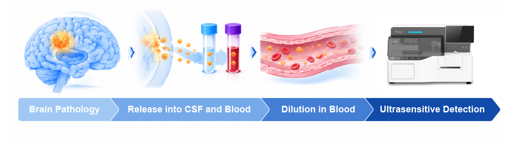
Total tau in blood is a limited readout of brain injury in chronic disease, because most tau measured in peripheral blood reflects non-brain sources. BD-tau assays address this by selectively detecting tau carrying the exon 4–5 junction, an isoform signature of CNS tau that peripheral "big tau" lacks. In validation cohorts, plasma BD-tau tracked CSF tau measures more closely than total tau did, and separated AD from other neurodegenerative diseases with greater accuracy[8]. BD-tau and its phosphorylated forms are a newer addition to the AD research toolkit; Vazyme's AD biomarker platform extends the tau panel beyond p-Tau181 and p-Tau217 to targets such as np-Tau217, p-Tau205, p-Tau231, and phosphorylated BD-tau forms(e.g. p-BD Tau217, p-BD Tau181), see the platform for the full target list.
● Phosphorylated tau biomarkers (p-Tau181 and p-Tau217)
p-Tau181 and p-Tau217 are tau phosphorylated at threonine 181 and threonine 217, respectively. Both rise along the AD continuum and reflect AD-related tau pathology. Research studies have reported strong performance for plasma p-Tau217 in distinguishing Alzheimer's disease from other neurodegenerative conditions, in several cohorts outperforming plasma p-Tau181, NfL, and other fluid measures[5]. However, performance estimates depend on the study population, reference standard, assay methodology, and predefined cutoff; published results describe the literature, not the performance of any single commercial kit. Plasma p-Tau181 was validated across four prospective cohorts and remains widely used, particularly where comparability with historical longitudinal data matters[6]. Vazyme's p-Tau217 (CLIA) research assay and p-Tau181 (CLIA) research assay sit within a broader AD portfolio that also covers Aβ1-40, Aβ1-42, GFAP, NfL, BD-tau, and additional tau-related targets. Supported sample types and species vary by biomarker and assay, consult the platform's sample compatibility matrix and the relevant product documentation before selecting an assay for plasma, CSF, tissue lysate/brain homogenate, or organoid culture supernatant studies.
● Amyloid-beta biomarkers (Aβ40 and Aβ42)
Aβ42 is the more aggregation-prone species and the main constituent of amyloid plaques; Aβ40 is more abundant overall but aggregates less readily. In human AD research, a lower Aβ42/40 ratio is commonly associated with cerebral amyloid pathology: as plaques accumulate, soluble Aβ42 falls relative to Aβ40. The magnitude and interpretation of this change depend on the sample matrix, assay methodology, and study population[2, 4]. Plasma Aβ is present at very low abundance, and reliable measurement required a generational leap in assay sensitivity: immunoprecipitation–mass spectrometry studies demonstrated that plasma Aβ42/40 and composite ratios predict brain amyloid-PET status with high accuracy[4]. Vazyme offers separate quantitative Aβ1-40 and Aβ1-42 (CLIA) research assays, but note that single-analyte assays are not automatically a validated ratio method. If you plan to calculate an Aβ42/40 ratio, verify analytical compatibility, measurement ranges, and the validity of the calculation for your study design.
● Glial fibrillary acidic protein (GFAP)
GFAP is an intermediate filament protein of astrocytes. Blood GFAP rises with astrocytic reactivity and correlates with amyloid pathology, making it a useful neuroinflammation readout in AD research, but it is not specific to AD, because astrocyte activation occurs in many brain disorders[7]. A dedicated GFAP (CLIA) research assay is available for this readout.
● Neurofilament light chain (NfL)
NfL is released from damaged axons and is a general marker of neuroaxonal injury. It is elevated in AD and rises with disease severity, but also in frontotemporal dementia, ALS, multiple sclerosis, and traumatic brain injury[7]. In AD studies, NfL is best interpreted as a severity, progression, and treatment-response marker rather than an AD-specific marker, a use case served by Vazyme's NfL (CLIA) research assay.
Core AD Biomarkers at a Glance
The table below summarizes the core AD biomarkers at a glance, the biological process each one reflects, and its typical research role.
Biomarker | What it reflects | Typical direction in AD | Common sample types | Primary research role |
Aβ42/40 ratio | Amyloid plaque aggregation (A) | Decreases as plaques accumulate | Plasma, CSF | Amyloid pathology readout; cohort stratification |
p-Tau181 | Tau phosphorylation (T) | Increases along the AD continuum | Plasma, CSF | AD vs. non-AD differentiation; longitudinal tracking |
p-Tau217 | Tau phosphorylation; tracks amyloid and tau pathology (A/T) | Rises early; closely tracks amyloid and tau burden | Plasma, CSF | Early-pathology detection research; staging |
GFAP | Astrocytic reactivity / neuroinflammation | Increases with amyloid pathology | Plasma, CSF | Neuroinflammation readout; progression |
NfL | Neuroaxonal injury (N) | Increases with injury severity | Plasma, CSF, serum | Severity and progression; treatment monitoring (not AD-specific) |
Total tau | General neuronal injury | Elevated in acute CNS injury | Plasma, CSF | Acute injury; limited specificity in chronic AD |
BD-tau | Brain-derived tau (N, AD-type) | Increases with AD pathology | Plasma, CSF | AD-type neurodegeneration readout with reduced peripheral interference |
Table 1. Core AD biomarkers at a glance
Directions are typical findings from human cohorts; values, and occasionally directions, can differ across species and disease models. Sample types listed are those commonly reported in the literature; the validated sample types for a specific Vazyme assay are specified in its instructions for use and on the platform's compatibility matrix. Confirm behavior in your own matrix using assay-specific validation data.
To see all of Vazyme's AD biomarkers at a glance, including the additional tau targets (np-Tau217, p-Tau205, p-Tau231, and phosphorylated BD-tau forms) and the sample types and species each assay supports, explore the Alzheimer's Biomarker Platform. If you need any help selecting assays for your study, contact our technical team.
What Sample Types Are Used in Alzheimer's Biomarker Research?
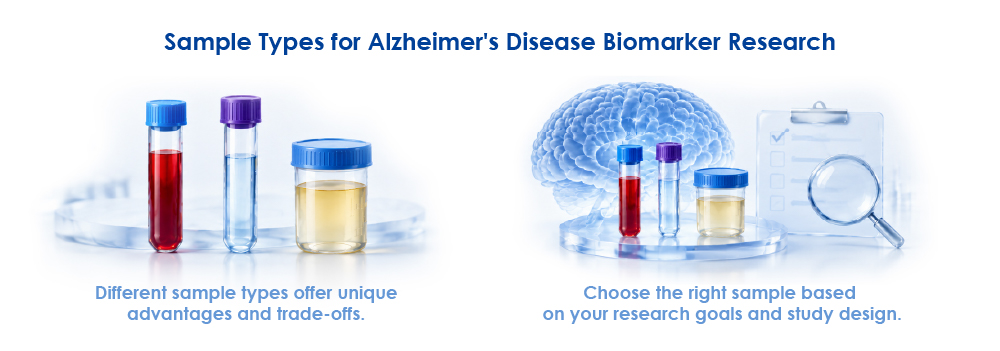
No single sample type is "best." The right matrix depends on your research question, your model, and your assay's validated compatibility.
●CSF is the established reference matrix for core AD biomarkers and sits closest to the brain interstitial fluid, but sampling is invasive, which limits large cohorts and repeated measures.
●Blood (plasma) is minimally invasive and supports large-cohort screening and longitudinal follow-up. The trade-off is concentration: early-pathology signals can be present at fg/mL levels, so plasma Aβ and p-tau assays must be ultrasensitive[3, 7].
●Plasma vs. serum: anticoagulant choice matters. EDTA plasma is the most common reference matrix in standardized plasma AD biomarker protocols, and collection tube type alone shifted measured levels of every assessed blood biomarker by more than 10% in a standardized evaluation[9].
●Tissue lysate and brain organoid culture supernatant support mechanistic and model studies where fluid biomarkers cannot capture spatial or cellular detail.
Pre-analytical handling is part of assay selection, not an afterthought. In the Global Biomarker Standardization Consortium's evidence-based protocol study, Aβ peptides were the most sensitive to processing delays (declining by more than 10%, and more steeply at room temperature), NfL and GFAP increased by more than 10% under room-temperature storage, and p-tau isoforms, particularly p-tau217, were the most robust across tube type, delay, centrifugation, and freeze–thaw variations[9]. The practical rule: standardize tube type, processing time, centrifugation, and freeze–thaw cycles within your study, and follow the assay's instructions for use.
How Are Alzheimer's Biomarkers Used in Research?
●Pathology and mechanism studies, quantifying amyloid, tau, inflammatory, and injury markers in human cohorts and relating them to clinical or imaging measures.
●Model characterization, transgenic models (amyloid models such as 5×FAD and APP/PS1; tau models such as P301S), non-human primates, and brain organoids. Marker behavior and concentration ranges differ from human samples, so species cross-reactivity and matrix compatibility must be validated per assay. Vazyme's platform documents validated sample-type combinations across human, mouse (WT/5×FAD/P301S), macaque, pig, and brain organoid matrices[3].
●Therapeutic development, target engagement, pharmacodynamic markers, and cohort stratification in preclinical and translational studies.
●Longitudinal monitoring, tracking progression or treatment response, where blood sampling enables repeated measures that CSF cannot.
A 2026 peer-reviewed evaluation of an automated chemiluminescent plasma assay (Vazyme AD Assay) measuring six AD biomarkers in a Chinese memory clinic cohort, 409 participants, with Aβ-PET as the reference standard, reported AUCs of 0.92–0.95 for p-tau217 and p-tau217-based ratios in detecting amyloid pathology, with consistent performance across freshly analyzed and frozen batch-processed samples[10]. A useful reminder that both the assay platform and the pre-analytical workflow shape the data you get, and that clinical study results do not change the research-use-only status of the assays.
How to Select Biomarkers for Your AD Study
A practical sequence for building your panel:
1.Define the research question. Pathology detection, intervention response, longitudinal progression, or model characterization? The question determines whether you need A, T, (N), or inflammation markers, or a combination.
2.Fix the sample matrix first. Plasma, CSF, tissue, or organoid supernatant, this constrains which assays are usable at your target concentrations.
3.Match markers to biology. Use AT(N) as the checklist: an amyloid readout (Aβ42/40), a tau readout (p-Tau181 or p-Tau217), and, where neurodegeneration is the endpoint, NfL, total tau, or BD-tau and its phosphorylated forms, plus GFAP if neuroinflammation is in scope.
4.Evaluate assay performance, not just marker names. Check the limit of detection against expected concentrations, linear range, precision (repeatability and intermediate precision), antibody specificity and cross-reactivity, and validated species and sample-type compatibility.
5.Plan comparability and validation. Decide early which published assays your data will be compared against, and build in QC samples and appropriate controls.
For a decision-oriented walkthrough, including how to read assay specifications, see our companion guide on choosing AD biomarker assays (link added at publication). To review the full target list, including additional research targets such as UCH-L1 and ApoE4, and the sample types and species covered, explore Vazyme's Alzheimer's Biomarker Platform.
Planning an AD biomarker study? Choosing the right biomarker depends on your research question, sample type, species, and assay requirements. Explore Vazyme's Alzheimer's Biomarker Platform to review the available biomarker targets and supported sample types, then tell us which biomarkers, samples, and research models you are working with, and our team will help you identify the relevant product documentation and suitable assay options.
Explore the Alzheimer's Biomarker Platform
Frequently Asked Questions
1.What are the main biomarkers of Alzheimer's disease?
The key biomarkers are amyloid-beta (Aβ40 and Aβ42, usually measured as the Aβ42/40 ratio), phosphorylated tau (p-Tau181 and p-Tau217), GFAP (astrocyte activation), NfL (neuroaxonal injury), and total tau or brain-derived tau (BD-tau) and its phosphorylated forms. The AT(N) framework groups them into amyloid (A), tau (T), and neurodegeneration (N) categories.
2.What is the difference between amyloid and tau biomarkers?
Amyloid biomarkers reflect Aβ plaque aggregation and typically become abnormal earliest, in AD, the CSF/plasma Aβ42/40 ratio falls as plaques accumulate. Tau biomarkers (p-Tau181, p-Tau217) reflect tau phosphorylation and tangle pathology, rise as the disease progresses, and track more closely with symptom severity and neurodegeneration.
3.Which biomarkers are studied in Alzheimer's disease mouse models?
Amyloid models such as 5×FAD and APP/PS1 are typically studied with Aβ42/40 measurements in plasma, CSF, or brain tissue; tau models such as P301S are studied with p-tau species. Absolute concentrations and effect directions can differ from human cohorts, so species-specific assay validation is essential before comparing across models.
4.Can blood-based biomarkers replace CSF or PET measurements in research?
They complement rather than replace them. Plasma assays are less invasive and scalable, and modern ultrasensitive assays show strong agreement with CSF and imaging measures, but CSF and imaging remain reference standards for many questions, and fluid and imaging biomarkers within the same category are not always interchangeable[2].
5.What sample types and species are supported by Vazyme's AD biomarker assays?
Vazyme's AD biomarker portfolio covers multiple biomarkers and research models, including human, mouse (WT/5×FAD/P301S), macaque, pig, and brain organoid matrices, with sample types spanning plasma and CSF to tissue lysate and organoid culture supernatant. Supported combinations vary by assay. Refer to the Alzheimer's Biomarker Platform compatibility matrix and the relevant product documentation to confirm whether a specific assay suits your sample type, species, and research objective.
6.Are Alzheimer's disease biomarkers used for diagnosis?
In clinical practice, AD diagnosis increasingly incorporates validated biomarker tests. The assays referenced in this article are for research use only and are not for use in diagnostic procedures[3].
References
1.Jack CR Jr, Bennett DA, Blennow K, et al. NIA-AA Research Framework: Toward a biological definition of Alzheimer's disease. Alzheimers Dement. 2018;14(4):535–562. doi:10.1016/j.jalz.2018.02.018
2.Jack CR Jr, Andrews SJ, Beach TG, et al. Revised criteria for diagnosis and staging of Alzheimer's disease: Alzheimer's Association Workgroup. Alzheimers Dement. 2024. doi:10.1002/alz.13859
3.Vazyme. Alzheimer's Biomarker Platform, p-Tau217, BD-Tau & chemiluminescence. Available at: info.vazymeglobal.com/alzheimer-biomarkers
4.Nakamura A, Kaneko N, Villemagne VL, et al. High performance plasma amyloid-β biomarkers for Alzheimer's disease. Nature. 2018;554(7691):249–254. doi:10.1038/nature25456
5.Palmqvist S, Janelidze S, Quiroz YT, et al. Discriminative accuracy of plasma phospho-tau217 for Alzheimer disease vs other neurodegenerative disorders. JAMA. 2020;324(8):772–781. doi:10.1001/jama.2020.12134
6.Karikari TK, Pascoal TA, Ashton NJ, et al. Blood phosphorylated tau 181 as a biomarker for Alzheimer's disease: a diagnostic performance and prediction modelling study using data from four prospective cohorts. Lancet Neurol. 2020;19(5):422–433. doi:10.1016/S1474-4422(20)30071-5
7.Teunissen CE, Verberk IMW, Thijssen EH, et al. Blood-based biomarkers for Alzheimer's disease: towards clinical implementation. Lancet Neurol. 2022;21(1):66–77. doi:10.1016/S1474-4422(21)00361-6
8.González-Ortiz F, Turton M, Kaur D, et al. Brain-derived tau: a novel blood-based biomarker for Alzheimer's disease-type neurodegeneration. Brain. 2023;146(3):1152–1165. doi:10.1093/brain/awac407
9.Verberk IMW, Gouda M, Antwi-Berko D, et al. Evidence-based standardized sample handling protocol for accurate blood-based Alzheimer's disease biomarker measurement: Results and consensus of the Global Biomarker Standardization Consortium. Alzheimers Dement. 2025. doi:10.1002/alz.70752
10.Chen S, Wang FY, Li R, et al. Diagnostic performance of an automated plasma p-tau217 chemiluminescent assay for detecting Aβ pathology in a Chinese memory clinic cohort. J Prev Alzheimers Dis. 2026. doi:10.1016/j.tjpad.2026.100613
For research use only.

